[Home][Help][About RsiteDB][Download files]

Abstract :The database details the interactions of extruded, unpaired RNA nucleotide bases. It presents and classifies the protein binding pockets that accommodate them. It allows the recognition of similar protein binding patters involved in interactions with different RNA molecules. Given an unbound structure of a target protein it allows the prediction of its RNA nucleotide binding sites.
Input : The user can specify a PDB code of interest or upload the structure of a target protein. The protein structure can be unbound. If the structure is uploaded it should be in a PDB file format.

Chain specification: The input structure is analyzed and the user is prompted to select the protein chain of interested. If all protein chains should be considered the user should select the "All chains" option, but this will slow down the computation.

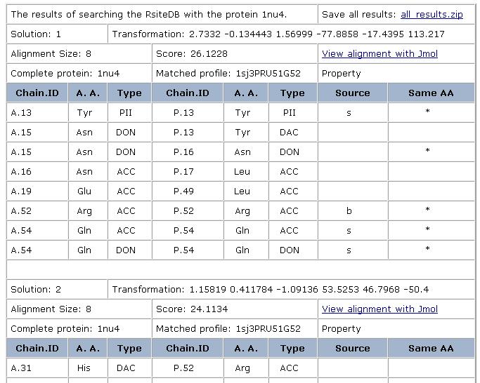
Prediction of dinucleotide binding sites: Here, we use the created clusters to predict RNA binding sites that accommodate unpaired extruded dinucleotides. Specifically, given a target protein structure not used for the classification, we search its surface for regions similar to the created 3D consensus binding patterns. These regions are predicted to serve as dinucleotide binding sites. Using leave-one-out tests, the success rate of these predictions was estimated to be about 80%. It must be noted that currently we do not aim to predict whether a protein can bind RNA; rather, given an unbound RNA binding protein, our goal is to predict its binding sites and their modes of interaction. In addition, due to a low number of single nucleotide clusters, currently, we do not use them for the prediction.
Given the target protein, we recognize and detail the regions that are similar to the created dinucleotide 3D consensus binding patterns. We detail the PDB code and the dinucleotides that were used to create the pattern as well as the alignment size, score and a transformation that can superimpose it on the target protein in 3D space. We provide a PDB file with this superimposition and allow its visualization with Jmol. In addition we detail the physico-chemical pattern that was found in the target protein. The first 3 columns provide the details of the target protein and the next two refer to the protein that originated the 3D consensus pattern. The following fields are presented: Chain.ID - the protein chain identifier and the amino acid PDB sequence number;
Amino Acid - The one letter amino acid code. However it must be noted that the method is based on the physico-chemical properties and does not consider the identity of the amino acids. These are only displayed for the convenience of analysis.
Type - The type of physico-chemical property that is matched by the algorithm. The method is based on a representation of each amino acid of a protein as a set of features that are important for its interaction with other molecules. The abbreviations of these features are: DON - Hydrogen bond donor; ACC - Hydrogen bond acceptor; DAC - Hydrogen bond donor and acceptor (e.g in histidine); ALI - Aliphatic Hydrophobic property; PII - Aromatic property (pi contacts).
Source This field specifies whether the matched property is contributed by the backbone or the side-chain of the amino acid. The abbreviations are: b - feature contributed by the backbone s - feature contributed by the backbone
Same AA: Marks the features shared by the two molecules that are contributed by residues with the same identity of the amino acid.

Each solution can be visualized with Jmol. The default view presents the superimposition of all the complexes, with their corresponding nucleotides/dinucleotides and the matched physico-chemical properties. The molecules are colored by model (according to the order of input molecules, the first is cyan, the second is magenta, the third is yellow). The physico-chemical properties (pseudocenters) are represented as balls. Hydrogen bond donors are blue, acceptors - red, donors/acceptors - green, hydrophobic aliphatic - orange and aromatic - gray. The surface points that have a similar location in all the molecules are represented as smaller dots with the same coloring.


Reference: Shulman-Peleg A, Shatsky M, Nussinov R. and Wolfson H. J. (2008) Prediction of interacting single-stranded RNA bases by protein binding patterns, Journal of Molecular Biology, Vol 379, pp 299-316.
Email:shulmana@tau.ac.il, ppdock@tau.ac.il